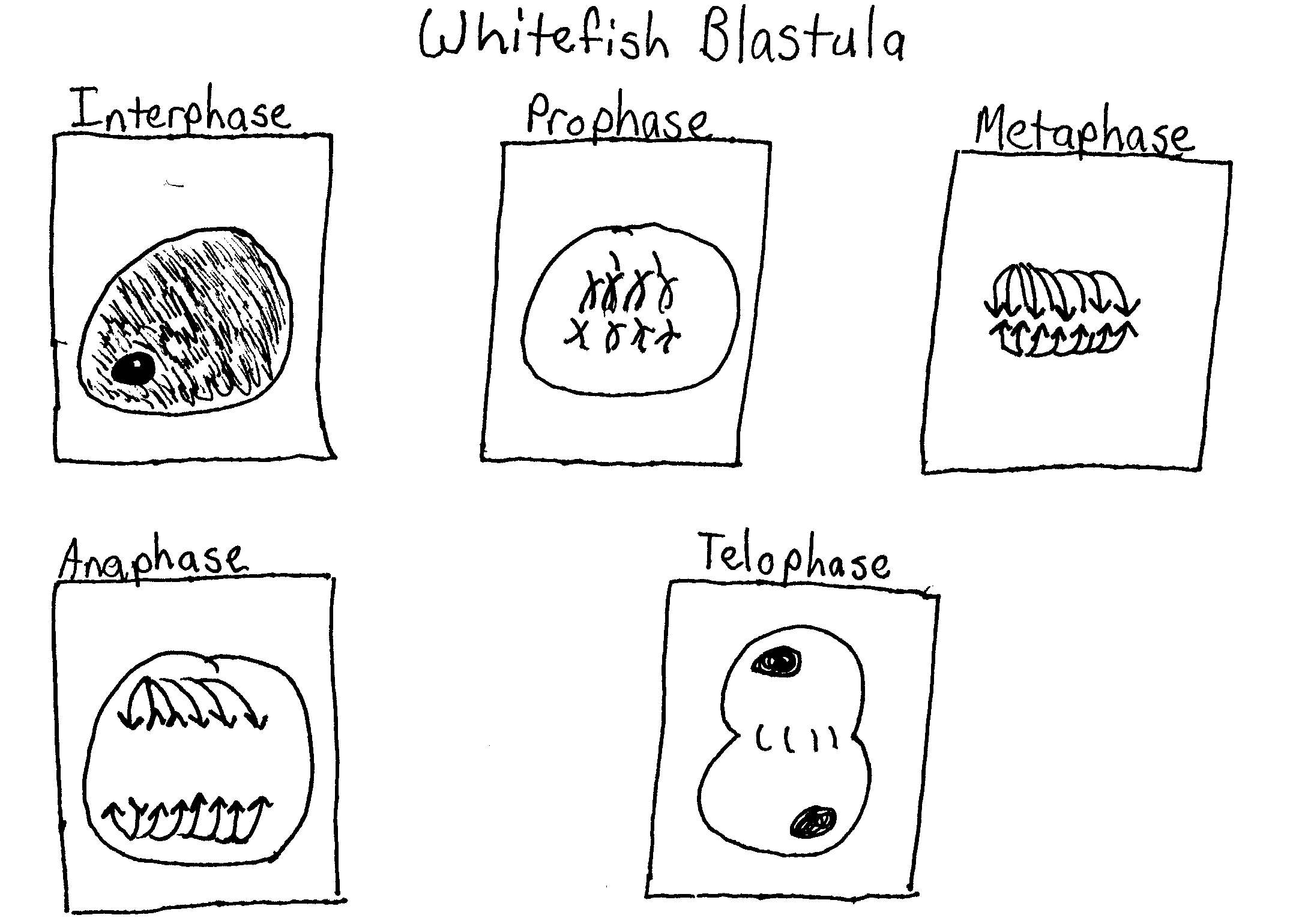
Whitefish Blastula Mitosis Drawing
Whitefish Blastula Mitosis Drawing - Web whitefish blastula cell mitosis. Observe the prepared slide of a whitefish blastula under high power (400x). Web the photomicrographs below show sections of whitefish blastula. Draw and label all stages of mitosis below. The whitefish embryo is a good place to look at mitosis because these cells are rapidly dividing as the fish embryo is growing. Web two specimens are commonly used by biologists to study mitosis: These undifferentiated cells undergo mitosis at a regular interval as the embryo increases in number of cells and complexity. Web the student will correctly identify and draw four stages of mitosis using microscope slide images of onion root tips and whitefish blastulae. Interphase, prophase, metaphase, anaphase, and telophase. List three reasons why organisms need to produce new cells.
Web since early embryogenesis involves rapid cellular division, the whitefish blastula has long served as a model of mitotic division in animals. Commercially available pop bead kits (e.g carolina biological supply company, item #171100) 40 pop beads of one color (red) Events & activities of each + slide of whitefish blastula slides learn with flashcards. Web whole mounts of whitefish blastula will illustrate reproductive cells in animals. The slides below show longitudinal sections of allium (onion) root tip. Nuclear membrane breaks down, chromatin condenses, mitotic spindle forms and attaches to kinetochores. Practice locating each of the stages of mitosis in the following photomicrographs.
The cell cycle is briefly described and broken down into mitosis, g1, s, and g2. Web whole mounts of whitefish blastula will illustrate reproductive cells in animals. Web the phases of mitosis. It also has the advantage of demonstrating clear spindle formation in the cytoplasm. The slides below show sections of whitefish blastula.
Lab & Ap Sample 2 Mitosis & Meiosis BIOLOGY JUNCTION
Mitotic cell division stages of Whitefish blastula YouTube

13th View of Whitefish Blastula with Mitosis Stages Labele… Flickr

Chapter 8 handout blks_ 10182011

Stages of Mitosis in the Blastula of a Whitefish Lab Manual for

AP Lab 3 Sample 3 Mitosis BIOLOGY JUNCTION

SOLVED Observe the stages of mitosis in the prepared slides of

Onion root tip whitefish blastula; The slides below show longitudinal sections of allium (onion) root tip. Web the student will correctly identify and draw four stages of mitosis using microscope slide images of onion root tips and whitefish blastulae. Because growth in roots occurs at the tips, this is where cells will most actively undergo mitosis. Web this series of images shows the progression of cells through prophase. Web whitefish blastula cell mitosis.
Web two specimens are commonly used by biologists to study mitosis: Obtain a whitefish blastula (early embryo) slide and find a cell in each of these phases: The blastula of a whitefish and the root tip of an onion. Web since early embryogenesis involves rapid cellular division, the whitefish blastula has long served as a model of mitotic division in animals. The cell cycle is briefly described and broken down into mitosis, g1, s, and g2.
These undifferentiated cells undergo mitosis at a regular interval as the embryo increases in number of cells and complexity. The slides below show sections of whitefish blastula. Commercially available pop bead kits (e.g carolina biological supply company, item #171100) 40 pop beads of one color (red) The blastula of a whitefish and the root tip of an onion.
Practice Locating Each Of The Stages Of Mitosis In The Following Photomicrographs.
The slides below show longitudinal sections of allium (onion) root tip. Observe the prepared slide of a whitefish blastula under high power (400x). To do the mitosis lab, you must have access to microscopes and have a set of mitosis slides. 4.4k views 1 year ago.
Web The Photomicrographs Below Show Sections Of Whitefish Blastula.
Hover over each image to learn about the specific events that occur during each mitotic phase. Web the cell is binucleate very briefly, mitosis ends. Examine the slide under a microscope. Web the phases of mitosis.
Identify And Describe The Stages Of Mitosis In Whitefish Blastula Cells.
Because growth in roots occurs at the tips, this is where cells will most actively undergo mitosis. Web the student will correctly identify and draw four stages of mitosis using microscope slide images of onion root tips and whitefish blastulae. You will make observational drawings and be prepared to take a practical quiz. Commercially available pop bead kits (e.g carolina biological supply company, item #171100) 40 pop beads of one color (red)
The Blastula Is An Early Stage Of Embryo Development, And Represents A Period In The Organism's Life When Most Of The Cells Are Dividing Consistently.
Web identify and draw a cell in each of the four stages of mitosis in the onion slide. Web whole mounts of whitefish blastula will illustrate reproductive cells in animals. Identify and draw a cell in each of the four stages of mitosis in the whitefish blastula slide. Web a blastula is a sphere of cells produced during the development of an embryo by repeated mitosis and cleavage of a fertilized egg.